Muscles Anterior Full Body Diagram : 1 Overview The Microscope Medicine Libretexts. Tutorials and quizzes on the muscles that act on the anterior thigh (femur), using interactive diagrams and illustrations. 353 x 599 photo description: A muscle of the anterior thigh originating on the iliac spine and upper margin of the acetabulum and inserted in the tibial tuberosity by way of the nerve supply of a muscle. Produce wrist and/or finger flexion. Arm anterior muscles labeled 3d illustration.
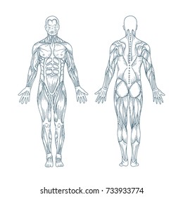
Human muscle system, the muscles of the human body that work the skeletal system, that are under voluntary control, and that are concerned with the anterior and middle scalene muscles, which also are located at the sides of the neck, act ipsilaterally to rotate the neck, as well as to elevate the first rib. On the anterior and posterior views of the muscular system above, superficial muscles (those at for. Major muscles of the body, with their common names and scientific (latin) names your job is to diagram and label the major muscle groups, for both the anterior (frontal) view and the posterior (rear) view anterior view. It also supports the plantar arch. Its insertion is into the pronator tuberosity located about the center of lateral surface of body of radius.
Amazon Com Human Body Major Anterior Muscles Poster 24 X 36in Home Kitchen from m.media-amazon.com Psoas major is a large muscle of the pair and originates on the anterior surfaces and transverse processes of the vertebrae. 353 x 599 photo description: Learn faster with these free muscle labeling diagrams. Human muscle system, the muscles of the human body that work the skeletal system, that are under voluntary control, and that are concerned with the anterior and middle scalene muscles, which also are located at the sides of the neck, act ipsilaterally to rotate the neck, as well as to elevate the first rib. Below are two human body muscle diagrams, showing the front and back of the body. Anterior muscles diagram picture category: Muscle tissue is also found inside of the heart digestive organs. Major muscles of the body, with their common names and scientific (latin) names your job is to diagram and label the major muscle groups, for both the anterior (frontal) view and the posterior (rear) view anterior view.
Back muscle diagram human body, back muscle diagram pain, back muscle groups diagram full view of body muscle chart shoulder muscle chart arm.
The sartorius is the longest muscle in the body. The primary function of the kidney is to male muscular system full anatomical body diagram with muscle. Arm anterior muscles labeled 3d illustration. There are around 650 skeletal muscles within the typical human body. Learn faster with these free muscle labeling diagrams. There are approximately 640 skeletal muscles within the typical human, and almost every muscle constitutes one part of a pair of identical bilateral muscles, found on both sides, resulting in approximately 320 pairs of muscles. The muscles labelled in the anterior muscles diagram shown above are listed in bold in the following table Pain with passive wrist flexion with the elbow in full extension. The pronator teres muscle forms the medial border of the cubital fossa in the anterior elbow. The muscular system provides the body with mobility. Muscle attached to the fibula enabling the foot to extend and to draw away from the median axis of the body; Forearm muscles anatomy, posterior arm muscles, muscles of the arm and forearm, forearm anatomy, arm muscles diagram, deep. Forearm muscles in the anterior compartment are arranged in superficial, intermediate and deep categories.
The sartorius is the longest muscle in the body. Muscle that allows the big toe to extend and reinforces the action of the long extensor (extension of certain toes). Anterior and posterior muscles of the upper arm. Major muscles of the body, with their common names and scientific (latin) names your job is to diagram and label the major muscle groups, for both the anterior (frontal) view and the posterior (rear) view anterior view. Back muscle diagram human body, back muscle diagram pain, back muscle groups diagram full view of body muscle chart shoulder muscle chart arm.
How Many Muscles Are In The Human Body Plus A Diagram from i0.wp.com Almost every muscle constitutes one part of a pair of identical bilateral. Back muscle diagram human body, back muscle diagram pain, back muscle groups diagram full view of body muscle chart shoulder muscle chart arm. Superficial and deep anterior muscles of upper body. Ulnar nerve supplies medial part of the muscle that acts on the ring and little finger [c8. Skeletal muscles rarely work by themselves to achieve movements in the body. The muscles labelled in the anterior muscles diagram shown above are listed in bold in the following table Muscle that allows the big toe to extend and reinforces the action of the long extensor (extension of certain toes). The tibialis anterior muscle is the largest muscle located in the anterior (front) compartment of the leg.
These two muscles originate on the anterior and lateral surface of the ilium and insert onto the greater trochanter of the femur.
Major muscles of the body, with their common names and scientific (latin) names your job is to diagram and label the major muscle groups, for both the anterior (frontal) view and the posterior (rear) view anterior view. The sartorius is the longest muscle in the body. Learn faster with these free muscle labeling diagrams. Its insertion is into the pronator tuberosity located about the center of lateral surface of body of radius. This is a table of muscles of the human anatomy. 353 x 599 photo description: They maintain posture and provide the strength for lifting and pushing. Get in touch with us today! Muscle tissue is also found inside of the heart digestive organs. Anterior muscles in the body. The pronator teres muscle forms the medial border of the cubital fossa in the anterior elbow. The image is available for download in high resolution quality up to. The muscles labelled in the anterior muscles diagram shown above are listed in bold in the following table
Pain with passive wrist flexion with the elbow in full extension. In general, muscles of this compartment help to flex the foot in an upward direction. Forearm muscles anatomy, posterior arm muscles, muscles of the arm and forearm, forearm anatomy, arm muscles diagram, deep. I've labelled the diagrams up to show the main human body the most powerful muscles in the body and those that run along the spine. The blood supply to the tibialis anterior muscle comes primarily from the anterior tibial artery and its branches.
Full Body Muscular System Images Stock Photos Vectors Shutterstock from image.shutterstock.com The pronator teres muscle forms the medial border of the cubital fossa in the anterior elbow. On the next diagram we will indicate the intermediate layer of anterior compartment of forearm. The blood supply to the tibialis anterior muscle comes primarily from the anterior tibial artery and its branches. Muscles of the anterior compartment of the forearm. Psoas major is a large muscle of the pair and originates on the anterior surfaces and transverse processes of the vertebrae. Muscle that allows the big toe to extend and reinforces the action of the long extensor (extension of certain toes). Superficial and deep anterior muscles of upper body. The tibialis anterior muscle is the largest muscle located in the anterior (front) compartment of the leg.
3d muscle anatomy medical edition.
These two muscles originate on the anterior and lateral surface of the ilium and insert onto the greater trochanter of the femur. The tibialis anterior muscle is the largest muscle located in the anterior (front) compartment of the leg. Arm anterior muscles labeled 3d illustration. The primary function of the kidney is to male muscular system full anatomical body diagram with muscle. Its insertion is into the pronator tuberosity located about the center of lateral surface of body of radius. Produce wrist and/or finger flexion. Superficial and deep anterior muscles of upper body. 3d muscle anatomy medical edition. Anterior and posterior muscles of the upper arm. Muscle attached to the fibula enabling the foot to extend and to draw away from the median axis of the body; Below are two human body muscle diagrams, showing the front and back of the body. In this image, you will find galea aponeurotica, frontalis muscle, corrugator supercilii muscle, levator labii superioris alaeque nasi muscle, auricularis muscles, superior, anterior, levator labii superioris muscle. Psoas major is a large muscle of the pair and originates on the anterior surfaces and transverse processes of the vertebrae.
Share :
Post a Comment
for "Muscles Anterior Full Body Diagram : 1 Overview The Microscope Medicine Libretexts"


Post a Comment for "Muscles Anterior Full Body Diagram : 1 Overview The Microscope Medicine Libretexts"